Media
CUHK and HKU Researchers Introduce 3D Printing Technology in Complex Cardiac Surgery Procedures
24 May 2016
A joint research team from The Chinese University of Hong Kong (CUHK) and the University of Hong Kong (HKU) is the first in Hong Kong to introduce three-dimensional (3D) printing technology to complex cardiac procedures for enhancing procedural efficacy and safety. Researchers from the Division of Cardiology of the Department of Medicine and Therapeutics, Faculty of Medicine, CUHK, and the Department of Mechanical Engineering, Faculty of Engineering, HKU, collaborated to use echocardiographic data, to create soft silicone-based models of complex cardiac structures using 3D printing. The models allow cardiologists to personalise planning for cardiovascular intervention for each patient. The practice was applied to a complex case of Left Atrial Appendage (LAA) occlusion last year and the patient is now in good condition. This case has been reported in the medical journal Circulation: Cardiovascular Interventions.
Facilitates formulation of operation strategies
The complexity and variability of the human heart structure pose certain challenges to interventional surgery procedures, such as repeated deployment attempts and incomplete seal, which may increase the risks of procedural complications and surgical failure.
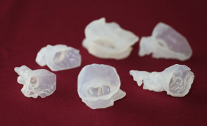
To overcome the challenges, researchers at the Division of Cardiology of the Department of Medicine and Therapeutics at CUHK applied 3D printing technology to plan complex cardiac procedures last year, the first time ever in Hong Kong. In collaboration with the Department of Mechanical Engineering of HKU, the team fabricated a silicone model of complex cardiac structures using 3D data from transesophageal echocardiography and 3D printing technology. With the 3D model, cardiologists can simulate the intervention procedures and formulate operation strategies in a more effective manner.
A 78-year-old female patient who was admitted for LAA occlusion to the Prince of Wales Hospital last year is one of the cases which benefited from the new technology. Transesophageal echocardiography revealed that the patient had a double-lobed LAA that would be challenging for occluder implantation as it is necessary to occlude the ostia of both lobes with a single device.
After simulating the procedures using a 3D silicone LAA model which was made to measure using the patient's transesophageal echocardiography data, cardiologists could implant the LAA closure device to the ideal position accurately. Hence, the risk of procedural complications or failure could be eliminated. Postoperative follow-up and assessment showed that the patient is in good condition.
Helps patients clearly understand procedures and facilitates training of cardiologists
Dr Alex Lee Pui-wai, Assistant Professor, Division of Cardiology, Department of Medicine and Therapeutics, Faculty of Medicine at CUHK, stated, ‘3D patient-specific models using materials that mechanically mimic soft cardiac tissues can enhance preoperative planning and appreciation of cardiac morphology by the doctors. This also helps patients better understand the operation procedures. The technology can be applied to other complex cardiac procedures and be used to train cardiologists as well.’
Dr Gary Cheung Shing-him, Clinical Assistant Professor (honorary), Division of Cardiology, Department of Medicine and Therapeutics, Faculty of Medicine at CUHK said, ‘Clinicians can reap the benefits of device simulation as 3D printing facilitates intervention procedures for cases with challenging cardiac structure anatomy.’
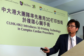
For the development of 3D printing technology in conjunction with clinical medicine, Dr Kwok Ka-wai, Assistant Professor, Department of Mechanical Engineering, Faculty of Engineering at HKU stated, ‘Advances in geometric modelling and 3D printing could add the cardiologist’s confidence in performing safer, more accurate and effective cardiovascular intervention procedures.’
Dr Alex Lee added that the team will further investigate the clinical applications of 3D printing technology in cardiology. Interested parties may e-mail to 3dprintingheart@cuhk.edu.hk for more information.
Media Enquiries:
Ms Esther Lau, Executive Officer (Development and External Relations), Faculty of Engineering, HKU (Tel: 3917 1924)
Ms Jackie Chan, Assistant Communications Manager, Faculty of Medicine, CUHK (Tel: 2632 4375)
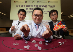
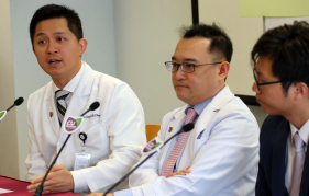
A joint research team from CUHK and HKU is the first in Hong Kong to introduce 3D printing technology to complex cardiac procedures for enhancing procedural efficacy and safety. (From left) Dr Gary Cheung, Clinical Assistant Professor (honorary), and Dr Alex Lee, Assistant Professor, from Division of Cardiology, Faculty of Medicine at CUHK; and Dr Kwok Ka-Wai, Assistant Professor, Faculty of Engineering at HKU.
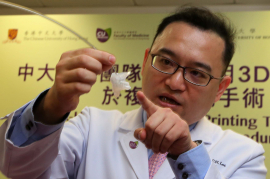
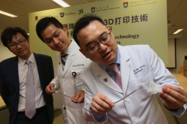
Dr Alex Lee, Assistant Professor, Division of Cardiology, Faculty of Medicine at CUHK states that 3D patient-specific cardiac models help patients better understand the operation procedures and enhance training of cardiologists.
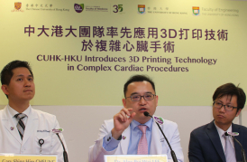
(Left) Dr Gary Cheung, Clinical Assistant Professor (honorary), Division of Cardiology, Faculty of Medicine at CUHK says 3D printing facilitates intervention procedures for cases with challenging cardiac structure anatomy.

