Media
Dinosaurs with laser beams on their heads
HKU’s Department of Earth Sciences: new laser-induced
fluorescence techniques uncover never-before-seen details in fossils
28 May 2015
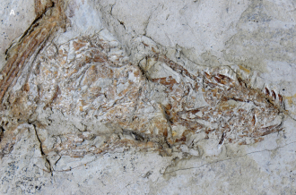
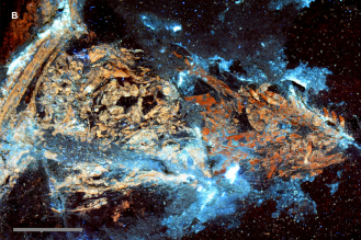
(The skull from specimen IVPP V13320 of the gliding carnivorous dinosaur Microraptor: is it a composite? A (left), the skull of IVPP V13320 under white light conditions shows subtle colour differences in the bone across a break in the rock slab – darker bone proximally and lighter bone distally. B (right), Under laser light stimulation, the bone fluoresces with the same colour pattern observed under white light conditions (see A) indicating that the colour differences relate to differences in the fossil’s mineral composition. The latter indicates that the skull is a composite specimen, but it is also possible - but less likely - that the pattern observed reflects variability in the conditions under which the animal was deposited and eventually transformed into rock. Scale bar 1 cm.)
Dr. Michael Pittman, Research Assistant Professor and head of the Department of Earth Science’s Vertebrate Palaeontology Laboratory, together with eight international colleagues have developed a simple new technique to analyse fossils. Published in the open-access journal “PLOS ONE”, the technique, called laser-stimulated fluorescence (LSF), utilises lasers to stimulate fluorescence in fossils that normally don’t fluoresce under standard UV lighting, with the new view photographed through a camera lens.
“Laser fluorescence opens the door to discovering previously unseen and unknown features in ancient fossils,” said Mr. Thomas Kaye, the study’s lead author and palaeontology research associate at the Burke Museum of Natural History and Culture, Seattle, USA.
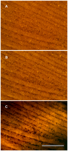
Each colour of laser emits a different wavelength of light, which excites the minerals that make up a fossil in different ways. LSF can be pinpointed onto microscopic fossils as well as projected over large areas to analyse bigger specimens. The research team applied this new technique to five different case studies in the “PLOS ONE” paper. One study involved fossilised feathers from the Green River Formation in western Wyoming. These feathers provide unusual insight into soft tissues preserved in the fossil record. When observed under standard lighting, barbs on the feathers are apparent but the interlocking barbules were missing. However, when back-lighting the fossils using the LSF technique, the much smaller barbules—become visible and were found to be widely distributed on the specimens. Multiple specimens from the same time and place showed these results. Without the new information provided by laser fluorescence, it might have been assumed that these feathers did not possess barbules.
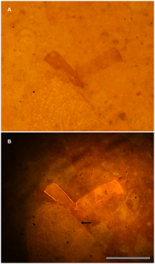
In addition to the fossilized feathers, the research team found that the LSF technique can identify specimens underneath the rock matrix the fossil is buried, help automate and speed up the sorting of micro-fossils from gravel, be used in the field to analyse fossils that can’t be removed from the landscape, and even identify possible faked or doctored “two-faced” fossils. For the latter case study the research team used a fossil of the gliding dinosaur Microraptor, a species of carnivorous dinosaur which are the animals that Dr. Pittman’s research focuses on.
A ~50 million year old bird feather from the Green River Formation, USA under different lighting conditions: A, White light, only barbs are visible. B, Polarized light, some traces of barbules. C, Laser-stimulated fluorescence of matrix behind the feather backlights the fossil, revealing the barbules in detail. Scale bar 0.5 mm.
Sub-surface imaging of fossils using laser-stimulated fluorescence: A, White light photograph shows the bone fragment on the right entombed within matrix. B, Specimen under laser fluorescence. Photograph shows a high level of detail invisible under white light. Note that another much larger fragment also becomes visible (arrow). Scale bar 0.5 mm
There is a short list of non-destructive techniques that are both affordable and accessible for use in palaeontology; LSF is a new addition to this list. It provides an instantaneous, non-invasive, geochemical fingerprint of fossilised bone, soft tissue, integument (protective layers like skin and plant cuticles) and the surrounding rock matrix. The ability to look for hidden specimens in a fossil’s rock matrix used to be only possible using X-rays, CT scans and other high-cost imaging methods. With LSF, researchers can set up a basic station quickly and for around HK$4000. It could mean many dinosaurs with laser beams on their heads, uncovering hidden fossils and a wealth of anatomical findings in the future.
Dr. Pittman added: “LSF [laser-stimulated fluorescence] promises to become an important mainstream palaeontological technique, so the team and I really hope that it fulfils its billing and helps to drive the field further forward.”
For press enquiry, please contact Ms Cindy Chan, Senior Communication Manager of Faculty of Science, at 3917-5286/ 6703- 0212 or by email at cindycst@hku.hk.
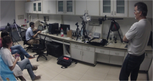
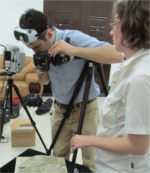
Research team members on a data collection trip to Beijing, China. Right: Thomas Kaye (lead author; Burke Museum of Natural History and Culture, USA); middle: Dr. Michael Pittman (Vertebrate Palaeontology Laboratory, HKU); left: Dr. Amanda Falk (Southwestern Oklahoma State University, USA).

